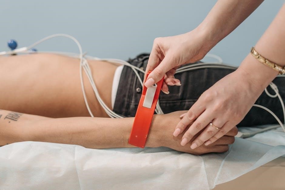
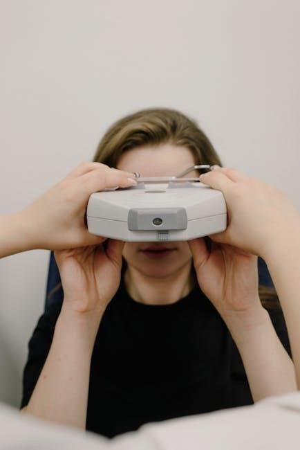
Electroencephalography (EEG) is a non-invasive neurophysiological test measuring brain activity, crucial for diagnosing various neurological conditions.
Precise electrode placement, following standardized systems like the 10-20 method, ensures reliable and interpretable data for accurate clinical assessments.
What is Electroencephalography (EEG)?
Electroencephalography (EEG) is a neurophysiological measurement technique used to record the electrical activity of the brain. It’s a non-invasive procedure, meaning it doesn’t require surgery or injection, making it a relatively safe and widely accessible diagnostic tool. EEG works by placing electrodes on the scalp to detect tiny electrical signals produced by the brain’s neurons. These signals are then amplified and displayed as wavy lines on a screen or recorded for later analysis.
The primary purpose of an EEG is to identify abnormalities in brain activity, which can be indicative of various neurological conditions. Understanding the fundamentals of EEG, and crucially, proper electrode placement according to systems like the 10-20 method, is paramount for accurate interpretation and effective clinical decision-making.
Importance of Accurate Electrode Placement
Accurate electrode placement is absolutely critical in electroencephalography (EEG) because even slight deviations from standardized positions – like those defined by the 10-20 system – can significantly impact the quality and interpretability of the recorded data. Misplaced electrodes can lead to inaccurate localization of brain activity, potentially resulting in misdiagnosis or ineffective treatment plans.
Consistent placement ensures that EEG recordings are comparable across different patients and studies. Following the 10-20 system allows neurologists to confidently correlate specific EEG patterns with underlying brain structures and functions. Standardized placement minimizes artifacts and noise, improving the signal-to-noise ratio and enhancing the reliability of the EEG findings. Therefore, meticulous attention to detail during electrode application is non-negotiable.

The 10-20 System: A Standard for EEG Electrode Placement
The 10-20 system provides standardized landmarks and measurements for consistent EEG electrode placement, ensuring reliable data and comparability across patients and studies.
Origins and Development of the 10-20 System
The 10-20 system emerged in the late 1950s, spearheaded by Herbert Jasper at the University of Montreal, aiming to standardize EEG electrode placement globally. Prior to this, variations in methodology hindered data comparison and interpretation. Jasper’s team sought a method based on readily identifiable skull landmarks, ensuring reproducibility.
The system’s name reflects its core principle: electrodes are placed 10% and 20% of the distance between key anatomical sites – nasion, inion, and preauricular points. This proportional approach allows for consistent placement regardless of head size or shape. Initial publications detailed the methodology, quickly gaining acceptance within the EEG community.
Over time, refinements and extensions have been added, but the fundamental principles remain unchanged, solidifying the 10-20 system as the gold standard for EEG electrode placement worldwide, facilitating reliable neurological assessments.
Key Anatomical Landmarks Used in the 10-20 System
The 10-20 system relies on three primary skull landmarks for accurate electrode placement: the nasion (the midpoint of the forehead between the eyes), the inion (the bony prominence at the back of the head), and the preauricular points (located just in front of the ears). These points serve as reference points for all measurements.
Measurements are taken between these landmarks to determine electrode positions. For example, 10% and 20% distances are marked along the midline from the nasion to the inion to locate central (Cz) and parietal (Pz) electrodes. Similarly, distances from the preauricular points define temporal electrode locations.
Consistent identification of these landmarks is crucial. Variations in landmark location can significantly impact EEG data. Careful palpation and, when necessary, anatomical references are essential for reliable and standardized electrode placement, ensuring accurate neurological interpretation.

Identifying Electrode Locations
EEG electrode locations are designated by letters representing brain lobes (F-frontal, C-central, T-temporal, P-parietal, O-occipital) and numbers indicating hemispheric placement.
Frontal Electrode Sites (Fp1, Fp2, F3, F4, F7, F8, Fz)
Frontal electrodes capture activity from the anterior portion of the brain, crucial for assessing executive functions, attention, and motor planning. Fp1 and Fp2 are prefrontal sites, positioned approximately 10% of the head length from the nasion (bridge of the nose) and equidistant from the midline.
F3 and F4 lie more centrally within the frontal lobe, while F7 and F8 are positioned more laterally, offering insights into orbitofrontal cortex activity. Fz represents the midline frontal site, providing a reference point for frontal lobe symmetry. These electrodes are vital for detecting frontal lobe abnormalities, including those associated with behavioral changes or frontal seizures. Accurate placement, guided by the 10-20 system, is paramount for reliable interpretation.
Central Electrode Sites (C3, C4, Cz)
Central electrodes are strategically positioned over the central sulcus, a critical brain region for processing somatosensory information and controlling voluntary movements. C3 typically records activity from the right side of the body, while C4 corresponds to the left side. This contralateral representation is fundamental to understanding motor cortex function.
Cz, the midline central electrode, serves as a crucial reference point for assessing symmetry in sensorimotor activity. These sites are particularly valuable in identifying focal abnormalities, such as those seen in stroke or seizure disorders affecting the motor or sensory cortices. Precise placement, adhering to the 10-20 system guidelines, ensures accurate localization of brain activity and reliable clinical interpretation.
Temporal Electrode Sites (T3, T4, T5, T6, T7, T8, Tz)
Temporal electrodes are positioned over the temporal lobes, vital for auditory processing, memory formation, and emotional regulation. T3 and T4 are located laterally, reflecting activity from the superior temporal gyri, while T5 and T6 are positioned more posteriorly, covering areas involved in auditory and visual integration.
T7 and T8 offer further lateral coverage, and Tz serves as the midline reference. These electrodes are crucial for identifying temporal lobe epilepsy, characterized by seizure activity originating in this region. Accurate placement, guided by the 10-20 system, is essential for differentiating temporal lobe seizures from other seizure types and for localizing the seizure focus for potential surgical intervention.
Parietal Electrode Sites (P3, P4, Pz)
Parietal electrodes, P3 and P4, are situated over the parietal lobes, key areas for spatial orientation, sensory integration, and navigation. They detect brain activity related to processing touch, temperature, pain, and pressure. P3 is typically placed on the left parietal region, while P4 corresponds to the right. These locations are vital for assessing somatosensory evoked potentials and identifying abnormalities related to parietal lobe dysfunction.
The midline electrode, Pz, provides a central reference point. These electrodes are particularly useful in diagnosing parietal lobe epilepsy, often manifesting as sensory disturbances. Precise placement, adhering to the 10-20 system guidelines, ensures accurate localization of seizure origins and aids in comprehensive neurological assessments.
Occipital Electrode Sites (O1, O2, Oz)
Occipital electrodes – O1, O2, and Oz – are positioned over the occipital lobes, the brain’s primary visual processing centers. O1 is located on the left occipital region, while O2 corresponds to the right. These electrodes are crucial for detecting brain activity associated with visual stimuli and assessing visual evoked potentials. They help identify abnormalities related to visual processing disorders or occipital lobe dysfunction.
The midline electrode, Oz, serves as a central reference point. These sites are particularly valuable in diagnosing occipital lobe epilepsy, which can present with visual hallucinations or disturbances. Accurate placement, guided by the 10-20 system, is essential for precise localization of seizure activity and comprehensive neurological evaluation.

Practical Considerations for Electrode Application
Skin preparation is vital for optimal signal quality; cleaning with abrasive gel reduces impedance. Electrode types and appropriate conductive paste ensure reliable recordings.
Skin Preparation Techniques
Effective skin preparation is paramount for acquiring high-quality EEG recordings, directly impacting impedance and signal clarity. The process begins with gentle cleansing of the scalp to remove oils, gels, and other contaminants that can impede electrical conductivity.
Subsequently, a mild abrasive agent, often a specialized EEG preparation gel or a gentle scrub, is applied to lightly exfoliate the skin. This crucial step removes dead skin cells, creating a more direct pathway for electrical signals.
Care must be taken to avoid excessive abrasion, which can cause skin irritation or micro-abrasions. Following abrasion, the skin should be cleaned again to remove any residual abrasive particles. Proper preparation significantly reduces impedance, minimizing noise and enhancing the accuracy of EEG data interpretation.
Electrode Types and Conductivity
EEG electrodes come in various types, including disc, cup, and saline-based electrodes, each offering unique advantages. Disc electrodes are commonly used for routine EEGs, while saline-based electrodes provide superior conductivity, particularly beneficial for high-density recordings. Cup electrodes offer versatility in application.
Conductivity, measured in impedance, is critical for signal quality. Lower impedance equates to better signal transmission. Electrode gels play a vital role in reducing impedance by filling the space between the scalp and the electrode, enhancing ionic conductivity.
Silver/silver chloride (Ag/AgCl) electrodes are preferred due to their stable and low-noise characteristics. Maintaining consistent conductivity across all electrodes is essential for accurate data interpretation and minimizing artifacts during EEG acquisition.
Wireless EEG Headsets & Emerging Technologies
Innovative wireless EEG headsets, like CerebAir and Imec’s prototypes, are revolutionizing brain monitoring, offering improved patient comfort and mobility during recordings.
CerebAir (Nihon Kohden) – Wireless ICU EEG
CerebAir, developed by Nihon Kohden, represents a significant advancement in continuous EEG monitoring for critically ill patients within intensive care units (ICUs). This wireless headset eliminates the constraints of traditional, wired EEG systems, allowing for greater patient mobility and reduced risk of cable-related complications. It functions as a telemetry EEG amplifier, utilizing eight electrodes to capture brainwave activity.
The system’s wireless nature is particularly beneficial in the ICU setting, where patient positioning and movement are frequent. CerebAir facilitates real-time monitoring of neurological function, aiding in the early detection of conditions like seizures, encephalopathy, and cerebral ischemia. Its ease of application and portability contribute to streamlined workflows for healthcare professionals, ultimately enhancing patient care and outcomes.
Imec/Holst Centre – Emotion & Cognitive Process EEG Headsets
Imec and Holst Centre have pioneered a novel EEG headset designed to measure intricate brain activity related to emotions and cognitive processes. This prototype represents a major leap forward in understanding the neural correlates of human experience, moving beyond simple diagnostic applications. The headset aims to decode complex mental states, offering potential for applications in areas like mental health monitoring and human-computer interaction.
This innovative technology utilizes advanced signal processing techniques to analyze EEG data, identifying patterns associated with specific emotional states and cognitive tasks. The development signifies a shift towards more sophisticated EEG applications, capable of providing insights into the subjective experience. It promises a future where brain-computer interfaces can respond dynamically to a user’s emotional and cognitive needs.

EEG in Epilepsy Diagnosis
EEG plays a vital role in epilepsy diagnosis, identifying seizure activity and characterizing the type of epileptic disorder. Analyzing interictal and ictal patterns aids precise localization.
Role of EEG in Identifying Seizure Activity
Electroencephalography (EEG) is a cornerstone in the diagnostic evaluation of epilepsy, primarily due to its ability to directly record electrical activity within the brain. Identifying seizure activity relies on recognizing characteristic patterns – often subtle – that deviate from normal brainwave patterns.
EEG can detect both ongoing seizure activity during an event (ictal patterns) and abnormal brainwave patterns between seizures (interictal patterns). The 10-20 system for electrode placement is crucial; it ensures consistent coverage and allows for accurate localization of seizure origins.
Specific seizure types manifest with distinct EEG signatures. For example, generalized spike-wave discharges are common in absence seizures, while focal slowing or sharp waves may indicate focal epilepsy. Careful interpretation, considering clinical context and precise electrode localization, is essential for accurate diagnosis and treatment planning.
Interictal and Ictal EEG Patterns
EEG patterns are categorized as either interictal – recorded between seizures – or ictal – recorded during a seizure. Interictal activity in epilepsy often reveals subtle abnormalities like spike-and-wave discharges, sharp waves, or focal slowing, indicating an increased predisposition to seizures. These patterns aren’t always present, necessitating prolonged monitoring.

Ictal patterns, conversely, are dramatically different. They exhibit rapid, evolving changes in frequency, amplitude, and morphology, often spreading across the scalp. The specific appearance depends on the seizure type and origin.

Accurate 10-20 system electrode placement is vital for capturing these patterns effectively. Proper localization helps pinpoint the seizure focus, guiding treatment decisions. Analyzing both interictal and ictal patterns provides a comprehensive understanding of the epileptic process, aiding in diagnosis and management.

Accessing 10-20 EEG Electrode Placement PDFs
Numerous online resources offer downloadable 10-20 EEG placement guides in PDF format, ensuring accessibility for clinicians and researchers needing accurate diagrams.
Online Resources for Downloadable PDFs
A wealth of online platforms provide readily accessible 10-20 EEG electrode placement PDFs, catering to diverse learning and practical needs. University websites, often within neurology or medical engineering departments, frequently host detailed guides for educational purposes. Professional organizations dedicated to EEG technology and clinical neurophysiology, such as the American Clinical Neurophysiology Society (ACNS), may offer resources to members or the public.
Searching using specific keywords like “10-20 system PDF,” “EEG electrode placement chart,” or “international 10-20 system” on search engines yields numerous results. It’s crucial to verify the source’s credibility before relying on the information. Many medical equipment suppliers also provide placement guides alongside their product offerings. Always prioritize resources from reputable institutions or organizations to ensure accuracy and adherence to established standards.
Reputable Sources for Accurate Placement Guides
Ensuring accuracy in 10-20 EEG electrode placement demands utilizing resources from highly trusted sources. The American Electroencephalographic Society (AEEG) stands as a premier authority, offering comprehensive guidelines and educational materials. Academic institutions with strong neuroscience programs, like Harvard Medical School and Johns Hopkins University, often publish detailed placement protocols and visual aids.
Medical textbooks specializing in clinical neurophysiology consistently feature accurate 10-20 system diagrams and explanations. Reputable medical publishers, such as Elsevier and Springer, are excellent starting points. Furthermore, manufacturers of EEG equipment, including Nihon Kohden and NeuroWave Systems, frequently provide detailed placement guides specific to their systems. Prioritizing these sources minimizes errors and promotes standardized, reliable EEG recordings for accurate diagnoses.